![]()
![]()
Deepwater Rice - investigations into the yellow stem borer in Bangladesh - 11 - 1982 research cont. |
A literature search showed that there was little published information on the anatomy of deepwater rice plants and no recorded study of the actual effect of feeding by S. incertulas larvae.
So, in a series of simple experiments, the bases of some 200 elongated stems of the variety Chamara collected during September and October 1982 were immersed in a weak solution of aniline blue dye for 2-3 days, The stems then were dissected and/or sectioned to investigate the effect of feeding by stem-borer larvae. The vascular tissue of the stems was clearly stained blue. Photographs were taken of several examples and drawings made from these are shown in Figs 1 & 2.
The photographs and other examples can be seen in detail in this linked page.
Figure 1
  | |||
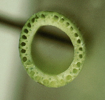 | |||
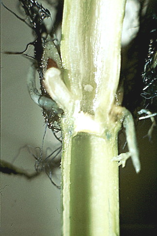
| The key structural characteristics of deepwater rice are clearly shown. In the middle elongated internodes there is a large central space, medullary cavity or lumen. The lumen is lined with soft ground tissue, the parenchyma. Between the parenchyma and the strong dermal layer, reinforced with fibrous, silicaceous tissue, is the denser sclerenchyma. Embedded within the sclerenchyma is a ring of air filled spaces, the aerenchyma. Also within the sclerenchyma, are an inner ring and an outer ring of vascular tissue, each ring being made up of bundles of xylem and phloem cells. In transverse view, it is the structure of the nodes which is of special interest. The centre of each node is filled with a septum and undifferentiated pith. The node itself is of dense reinforced tissue. The vascular tissue appears to form a broad continuous ring, into which run and leave the bundles seen in the longitudinal section. This ring also conveys the capacity to maintain nutrient flow evenly. | |||
| Figure 2 | |||
|  | ||
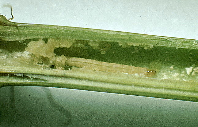
The longitudinal section, complete with in situ YSB larva, shows how in an elongation-stage internode the larva feeds primarily on the soft parenchyma, which is present in abundance. Moreover, as can be seen in the upper scelerenchyma,the two levels of conducting tissue (inner and outer, visible as blue lines) will maintain nutrient flow even if the inner level is fed upon.
![]() 1982 research cont. - more on DWR anatomy and YSB
1982 research cont. - more on DWR anatomy and YSB
| ©2000 - Brian Taylor CBiol FIBiol
FRES 11, Grazingfield, Wilford, Nottingham, NG11 7FN, U.K. Visiting Academic in the Department of Life Science, University of Nottingham |
href="\dwr\ysb10.htm"